 Cellosophy
Concept C1 · Strand C, Sensing and action
Receptors and signal transduction
Read first: Noise and limits of molecular sensing, Boundary, metabolism, and self-production
A bacterium has no eyes and no tongue, and it still knows when a few molecules of food have turned up. The knowing is done by proteins in its inner membrane, which turn one molecular bump into a change in what an enzyme is doing inside the cell. This page follows that conversion in Escherichia coli, the best-studied case by a mile, and asks two things: where does the signal get bigger, and where does chance sneak in?
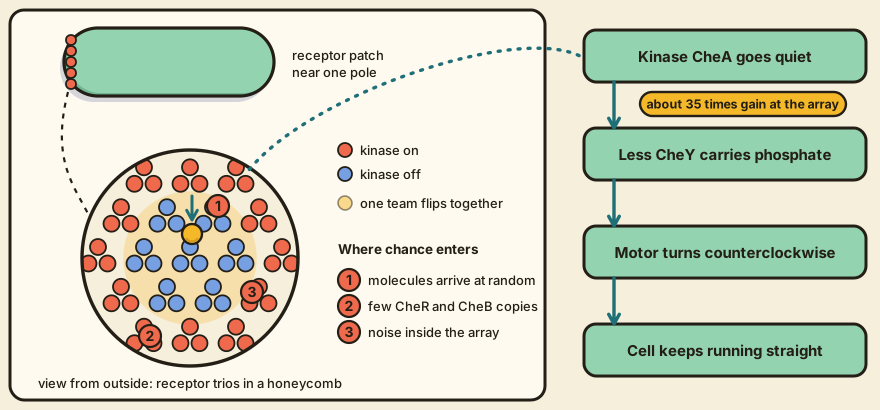
A molecule docks, a kinase calms down
E. coli has five kinds of chemoreceptor, called Tar, Tsr, Trg, Tap and Aer, and each responds to its own set of molecules (Muok et al., 2024). A receptor has a part that sticks out into the periplasm and grabs the chemical, helices that cross the membrane, and a part inside the cell that controls a kinase called CheA. When an attractant docks, the change in shape runs down the length of the receptor and turns CheA down. observed
A quieter CheA hands fewer phosphate groups to the messenger protein CheY. Phosphorylated CheY is what pushes the flagellar motor into clockwise rotation, which makes the cell tumble, and a phosphatase called CheZ strips the phosphate off fast, so the level of phosphorylated CheY tracks the kinase closely (Hazelbauer et al., 2008). Less of it, and the motors turn counterclockwise more often, and the cell keeps running straight. observed Food arrives, the cell stops changing its mind. That's the whole story in one line.
Receptors that only work in a crowd
The receptors don't sit alone. Cryo-electron tomography of whole cells shows patches of tightly packed receptors, with a layer of CheA and CheW at their inner tips, and most patches lie at a cell pole (Hazelbauer et al., 2008). Later structural work found that receptors form trimers of dimers, that these sit in a hexagonal lattice joined by CheA and CheW, and that the smallest unit able to signal is probably one CheA dimer with two CheW proteins and two receptor trimers (Muok et al., 2024). observed
How one molecule gets loud
Sourjik and Berg (2002) measured kinase activity in living cells through a fluorescence readout of how CheY with phosphate meets CheZ. In cells that can't methylate their receptors, half of the maximum response needed an attractant concentration equal to the receptor's dissociation constant. In normal cells it needed a concentration 35 times lower. observed Hazelbauer and colleagues describe about 36-fold of this gain as arising at the signalling complex, and at least 50-fold in the whole pathway, since a 1 percent change in receptor occupancy changes the motor's bias by 50 percent. Sourjik and Wingreen (2012) put the whole pathway nearer 100-fold, with the motor's switch supplying much of the rest.
In many signalling systems the gain comes from one activated enzyme churning out many product molecules. That trick isn't available here, because attractant binding lowers kinase activity, so a 36-fold gain means one occupied receptor has to sway about three dozen kinases, and the receptors have to be linked (Hazelbauer et al., 2008). The usual picture is a team of a few receptor trimers that flip together between a kinase-on and a kinase-off state, a picture drawn from models fitted to such data (Sourjik and Wingreen, 2012). modeled
We don't know how a change in receptor packing alters CheA activity, a gap Muok and colleagues state plainly. The size of the cooperating unit is also unsettled, since it could be one trimer or several trimers joined through shared CheA and CheW, and the arrays probably have holes in them (Hazelbauer et al., 2008).
Three doors for chance
Chance comes in through three doors. The first is outside the cell, because molecules arrive at the receptors at random, and Berg and Purcell (1977) showed that the precision of a concentration estimate is limited by that counting error. modeled The second is inside. CheR and CheB, the enzymes that reset the receptors, number only 200 to 400 copies per cell against about 15,000 receptors, so their levels vary from cell to cell. A review reports that this variation produces differences in adaptation time and a slow wandering of the motor's bias over tens of seconds (Sourjik and Wingreen, 2012). observed Because the adaptation system can't tell a spontaneous change in receptor activity from a stimulated one, fluctuation and response come bundled together. The adaptation loop page shows that mechanism.
The array itself is the third door. A gain of 35 or so applies to whatever reaches the array, so we infer that noise arising at the receptors gets magnified along with the signal. inferred An amplifier doesn't check what it's amplifying.
Which of the three doors lets in the most noise?
That's a live question. Mattingly and colleagues (2026) measured the information in single cells' kinase activity and compared it with the limit set by molecule arrival. They found that E. coli encode about two orders of magnitude less information than that limit, and concluded that noise in the cell's own signal processing, and not the arrival of molecules, is what holds it back. observed That's a challenge to an earlier claim, made without direct evidence, that arrival noise sets the limit.
Everything above is for E. coli. Muok and colleagues show that other bacteria build arrays with different symmetry and with extra adaptation systems, so these numbers shouldn't be carried over to them.
What you should be able to do after reading
Trace how an encounter outside the cell changes an activity inside it, naming the sources of amplification and noise along the way.
A quick test: take one attractant molecule landing on a Tar receptor and write out the next five steps, each as a change in a named protein, ending at the motor. Mark the step where one event becomes many, and the three places where chance gets in. C3 shows what the motor does with this signal, and C5 asks how the receptors reset.
You need first
References
- Hazelbauer, G. L., Falke, J. J., & Parkinson, J. S. (2008). Bacterial chemoreceptors: high-performance signaling in networked arrays. Trends in Biochemical Sciences, 33(1), 9-19. doi:10.1016/j.tibs.2007.09.014
- Muok, A. R., Olsthoorn, F. A., & Briegel, A. (2024). Unpacking alternative features of the bacterial chemotaxis system. Annual Review of Microbiology, 78, 169-189. doi:10.1146/annurev-micro-032421-110850
- Sourjik, V., & Berg, H. C. (2002). Receptor sensitivity in bacterial chemotaxis. Proceedings of the National Academy of Sciences, 99, 123-127. doi:10.1073/pnas.011589998
- Sourjik, V., & Wingreen, N. S. (2012). Responding to chemical gradients: bacterial chemotaxis. Current Opinion in Cell Biology, 24, 262-268. doi:10.1016/j.ceb.2011.11.008
- Berg, H. C., & Purcell, E. M. (1977). Physics of chemoreception. Biophysical Journal, 20(2), 193-219. doi:10.1016/S0006-3495(77)85544-6
- Mattingly, H. H., Kamino, K., Ong, J., Kottou, R., Emonet, T., & Machta, B. B. (2026). E. coli chemosensing accuracy is not limited by stochastic molecule arrivals. Nature Physics, 22(1), 123-130. doi:10.1038/s41567-025-03111-4