 Cellosophy
Pattern · complex scale
Receptor cluster: a crowd of receptors that acts as one switch
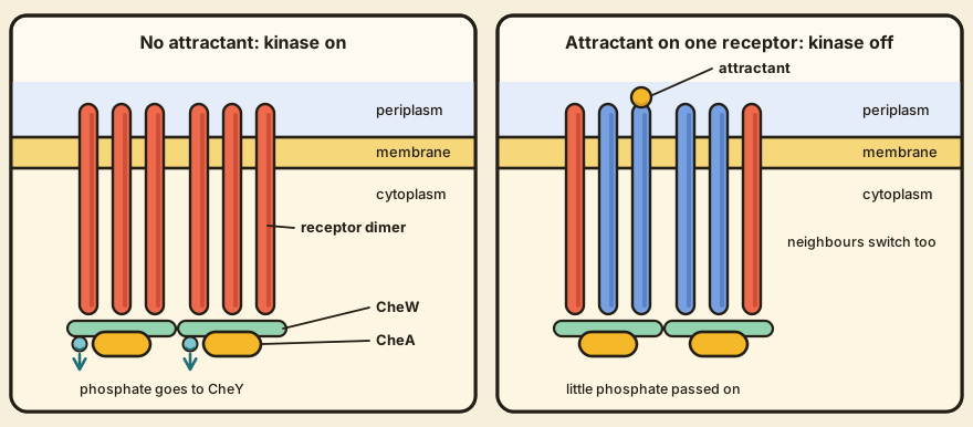
A bacterial chemoreceptor does its work as part of a crowd, and the crowd is what makes the cell so sensitive. In E. coli the receptors sit in dense patches, with the kinase CheA and its partner protein CheW packed against their inner ends, and a patch behaves like one switch that a few bound molecules can throw. Picture a stadium wave, with a handful of fans setting off a whole stand. This page looks at the patch at the scale of a complex, somewhere between a single receptor about 30 nanometres long and a whole patch about 250 nanometres across.
The problem: one molecule makes a very small noise
One receptor binding one molecule gives a very small signal, and the cell needs to react to small changes in concentration across a wide range. The size of the amplification has been measured. Hazelbauer, Falke and Parkinson (2008) report that the pathway amplifies a stimulus at least 50-fold, since a 1 percent change in receptor occupancy changes the rotation bias of the flagellar motors by 50 percent. observed
Most of that gain happens right at the receptors. Sourjik and Berg (2002) followed kinase activity in living cells with a fluorescence energy transfer method, and the review credits their result with 36-fold gain at the signalling complex. observed The usual story of gain in signalling is one activated enzyme churning out many product molecules, and in many systems that story is right. It can't be the story here, because attractant lowers the kinase's activity, so 36-fold gain means that one occupied receptor can control about three dozen kinases, and that implies a network linking each receptor to many copies of CheA. inferred We infer that receptors working alone couldn't do this, because each could then change only the kinase it touches.
How it works: needles, a floor, and a piston
A chemoreceptor is a needle-shaped dimer, about 300 angstroms long, standing roughly upright in the inner membrane. The end that sticks out into the periplasm binds the chemical. The end inside the cell can switch a kinase, and the review describes three modules in between: one that senses across the membrane, one that converts the signal, and one that controls the kinase. observed Cryo-electron tomography of intact cells shows the receptors packed side by side like wheat in a field, with a thin layer of CheA and CheW across their inner tips. Immunogold labelling confirms that the striations in the images are receptors and that the layer contains CheA and CheW, and patches fail to form when any one of the three proteins is missing. observed
The patch is a two-state switch. In the kinase-on state the complex raises the rate at which CheA phosphorylates itself several hundred-fold, and in the kinase-off state it turns that rate down. observed Attractant binding shoves a helix in the receptor towards the cytoplasm like a piston, and that shifts the complex towards kinase-off. observed CheA hands its phosphate to CheY, the messenger that reaches the motor, so what the cluster puts out is a flow of phosphate, which the signalling pathway carries on.
The cluster has a second input, and it pushes the other way. Methyl groups get added to the receptors at four to six sites on the inner part of the needle. Methylation moves the same helix in the opposite direction and favours kinase-on, and the kinase-off state is the one that's easier to methylate. observed This is how the adaptation loop resets the cluster: the methyl groups change what the same ligand does to the same receptors. Same key, same lock, a different click.
Why a crowd, when one receptor looks simpler
Experiments with receptors in nanodiscs, small plugs of membrane about 10 nanometres wide, show what one dimer can and can't do. Boldog and colleagues (2007), as the review describes them, put different numbers of dimers into each disc. A dimer alone bound ligand, got methylated, and got methylated faster when ligand was present. What it couldn't do was switch on CheA. Kinase activation peaked at an average of nearly three dimers per disc, and ligand shut it off just as in a cell. observed The results fit a trimer of dimers as the smallest unit that can run the kinase, though they can't rule out other sizes.
Trimers of dimers have also been seen directly. A crystal of most of the inner part of the serine receptor Tsr showed three dimers meeting at their tips, and cross-linking tests in living cells support the same arrangement. The contact residues are the same in all five E. coli receptors, so Tsr and Tar can sit in one mixed trimer, in proportion to how common each is. observed A committee of three, open to all comers.
The trimer is only the ground floor. The review reports that the response to attractant is cooperative, with Hill coefficients as high as 10, and that the presence of other receptor types, or of receptors at different methylation levels, changes the sensitivity. A Hill coefficient measures how steeply a response rises with its input, and a value of 1 would mean that every receptor acts alone. The authors read these results as an allosteric array with as many as several dozen receptors in the cooperating unit. inferred Patches are big, by the standards of a cell. The review gives an average diameter of about 250 nanometres, and says a circular patch of that size could hold 40 percent of the 7,500 receptor dimers in a cell while covering about 1 percent of its surface. Cells have no more than a few patches, and about 80 percent of patches sit at a pole. observed
What's still open: how the crowd talks to itself
We don't know how a conformational change in one receptor reaches the others. The review states that signalling within one dimer involves distinct changes in all three of its modules, and that how these change the trimer isn't yet fully defined. The size of the smallest working unit is open too, since it could be one trimer or several trimers linked through shared CheA and CheW, and the arrays probably contain gaps that fix the effective size of the cooperating group. inferred The same review says that how thousands of receptors come to gather in patches isn't understood. CheW and CheA compete for places on the receptor, and too much CheW gets in the way of trimer formation, so the proportions of the three proteins matter for the structure. How a cell sets them isn't something we found in the review.
Patches have turned up in all nine phylogenetically distant species examined, but there's little evidence on whether their receptors are also built from trimers, and a crystal of one archaeal receptor fragment showed rows of parallel dimers. Everything above is for E. coli, which is one bug among a great many.
That brings us to the question that gives the pattern its interest.
Should a receptor patch count as a sense organ, or only as a sensitive molecular switch?
We don't know. The evidence reviewed here shows a large gain and strong cooperation, which a switch needs, and it says nothing about whether a patch does anything beyond turning one chemical signal into a kinase signal. The concept page on receptors follows the same signal on into the cell.
Its parts
Chemoreceptor dimer (page to come): binds ligand outside the cell and sets the kinase on or off
CheW (page to come): couples the receptors to the kinase
CheA kinase (page to come): histidine kinase whose activity carries the signal
Wholes above it
References
- Hazelbauer, G. L., Falke, J. J., & Parkinson, J. S. (2008). Bacterial chemoreceptors: high-performance signaling in networked arrays. Trends in Biochemical Sciences, 33(1), 9-19. doi:10.1016/j.tibs.2007.09.014
- Sourjik, V., & Berg, H. C. (2002). Receptor sensitivity in bacterial chemotaxis. Proceedings of the National Academy of Sciences, 99, 123-127. doi:10.1073/pnas.011589998