 Cellosophy
Pattern · organelle scale
Flagellar motor: a proton-driven rotary switch
Set in the wall of a bacterium is a rotary motor about 45 nanometres across, spinning a long helical propeller and every so often throwing itself into reverse. The flagellar motor is the part of the cell that turns chemical information into a change of direction. Protons running down the cell's electrochemical gradient make it turn, and a small signalling protein decides which way. This page follows that arrangement in Escherichia coli, the cell most of what we know comes from.
The problem: no coasting, and no steering wheel
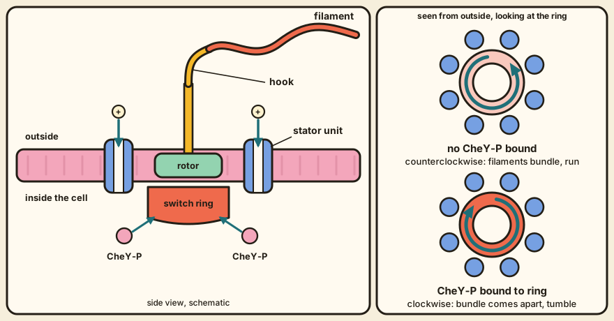
A bacterium swims through a fluid where it can't coast, so it has to keep driving a propeller just to keep moving. It also has to change course, because a cell that only swam straight would drift away from food as easily as toward it. Berg (2003) describes the E. coli answer. Each motor spins counterclockwise most of the time, and when every motor on the cell does so the filaments form a bundle that pushes the cell steadily forward. When one or more motors switch to clockwise, their filaments leave the bundle, and the cell tumbles and sets off in a new direction observed. Micali and Endres (2016) say a cell carries five to eight such motors, which is the number the rest of this page has in mind.
So one machine has two jobs. It has to turn with enough torque to move a cell, and it has to reverse fast when the chemotaxis system says so - engine and gearbox in one part. The chemotaxis module sits above this pattern and produces the signal. The signalling pathway delivers it, as phosphorylated CheY, which we write CheY-P.
How it works: a rotor, its stators, and a ring that listens
For this story the motor has three parts that matter, and they sit in the cell envelope in a fixed order. At the centre is the rotor, a set of rings that spin together. The lowest of them, inside the cell, is the switch ring built from the proteins FliG, FliM and FliN. Around the rotor in the inner membrane sit the stator units, made of the proteins MotA and MotB. A rod, a flexible hook and the filament carry the rotation outside observed. Berg counts about 20 kinds of part in all and Guo and Liu (2022) count about 25 proteins, a gap that probably reflects what each of them counts as a part. Biologists, like accountants, can disagree about categories.
Energy comes from the gradient across the membrane. In E. coli ATP plays no role in the turning itself, and the work is done by protons crossing through the stator units (Berg, 2003) observed. The speed is on the order of 100 turns per second, and Berg's torque-speed curve for E. coli crosses zero near 300 turns per second observed.
The stator unit's description has changed in recent years. Berg (2003) wrote that each unit was likely four MotA and two MotB. Then Santiveri and colleagues (2020) determined structures at about 3 angstroms by cryo-electron microscopy and found a pentamer of MotA around a dimer of MotB, a 5 to 2 ratio that they saw in proteins from Campylobacter, Shewanella and Vibrio observed. They didn't solve an E. coli stator, and they checked their structure against earlier mutation and cross-linking data from E. coli observed. Their model says that a proton binding to and leaving an aspartate on MotB makes the MotA pentamer rotate around the MotB dimer, and that the pentamer in turn pushes on the rotor protein FliG modeled. Guo and Liu (2022) call this a shift to seeing the stators as small rotary motors in their own right. Motors inside the motor, then.
Oddly, the number of stator units on one motor is less settled than the structure of one unit. Berg (2003) reviews experiments in which a paralysed motor was repaired by supplying a working gene, and its speed climbed in equal steps, up to eight and never nine observed. Freeze-fracture pictures show 10 to 12 studs in E. coli, and Berg suggests that two studs may make one torque generator, which would fit eight units inferred. Guo and Liu (2022) say roughly twelve stator units fit around the E. coli ring, and they report that units leave a motor faster under low load than under high load inferred. We don't know how many are working on a given motor at a given moment.
The switch is where it gets interesting. In Berg's account (2003), no known mutation in MotA or MotB damages switching alone, while mutations in FliG, FliM and FliN do, and CheY-P binds FliM observed. Santiveri and colleagues take this to mean that the signal acts on the rotor and that the same stator motion can drive either direction. Their proposal is that a turn of about 180 degrees in part of FliG lets it meet the other face of the MotA pentamer modeled. Guo and Liu (2022) report cryo-electron tomography of Vibrio and Borrelia motors in which the ring changes shape when CheY-P binds observed.
Switching is quick and sharp. In tethered E. coli cells the reversal takes no more than 10 milliseconds, and in single filaments it looks complete within 1 millisecond. Berg (2003) notes that one never sees a motor step through intermediate speeds observed. No dithering. Berg also reviews a picture in which the ring sits in one of two energy wells and CheY-P tilts the balance. Extrapolating from experiments at low temperature, he puts the difference between the two states at 14.4 kT at room temperature without CheY-P. In one analysis each bound molecule of an active CheY mutant lowered it by about 0.8 kT modeled.
The response is very steep. Cluzel and colleagues measured a Hill coefficient of 10.3 in single cells, as Berg (2003) reports. Yuan and Berg (2013) repeated the measurement on motors with a fixed number of FliM and found 16.5 and 20.7 for two groups of motors observed. They also report that the motor adjusts the number of FliM it holds as the steady level of CheY-P changes, which shifts the range where it's sensitive observed. The switch, in other words, can move where it listens.
What's still open: a decision, or a loaded coin
The binding of CheY-P to FliM is much less steep than the motor's response, and that gap is the puzzle. Berg (2003) quotes a Hill coefficient of 1.7 for binding, and Yuan and Berg (2013) say that if no higher binding cooperativity turns up, switching may need a non-equilibrium explanation inferred. We don't know which it is.
That leaves the question this pattern was chosen to raise.
A switch that reads one chemical message could be making a decision, or it could be a coin flip whose odds the message shifts. Which is it?
The two-well picture reads like the second, and the all-or-none reversals read like the first, though that's Claude's reading of these papers. Berg's thermal picture fits the exponential waiting times he reviews, and it leaves the choice between the two descriptions open. We don't know of a paper in this set that settles it, so for now the motor keeps its secret.
Its parts
Rotor and switch ring (page to come): FliG, FliM and FliN; the ring that CheY-P binds, which sets the direction of rotation
Stator unit (page to come): MotA and MotB, a proton channel that pushes on the rotor and so makes torque
Hook and filament (page to come): pass the rotation on to a helical propeller outside the cell
References
- Berg, H. C. (2003). The rotary motor of bacterial flagella. Annual Review of Biochemistry, 72, 19-54. doi:10.1146/annurev.biochem.72.121801.161737
- Santiveri, M., Roa-Eguiara, A., Kuhne, C., Wadhwa, N., Hu, H., Berg, H. C., Erhardt, M., & Taylor, N. M. I. (2020). Structure and function of stator units of the bacterial flagellar motor. Cell, 183(1), 244-257. doi:10.1016/j.cell.2020.08.016
- Guo, S., & Liu, J. (2022). The bacterial flagellar motor: insights into torque generation, rotational switching, and mechanosensing. Frontiers in Microbiology, 13, 911114. doi:10.3389/fmicb.2022.911114
- Yuan, J., & Berg, H. C. (2013). Ultrasensitivity of an adaptive bacterial motor. Journal of Molecular Biology, 425(11), 1760-1764. doi:10.1016/j.jmb.2013.02.016
- Micali, G., & Endres, R. G. (2016). Bacterial chemotaxis: information processing, thermodynamics, and behavior. Current Opinion in Microbiology, 30, 8-15. doi:10.1016/j.mib.2015.12.001